What is Electron Microscopy?
What is
Electron Microscopy? Examples, Principles, and Types
There's more to this world than
meets the eye... Literally!
Invisible to the human eye and
waiting to be discovered, there are so many complex hidden structures.
Fortunately, with the power of electron microscopy, scientists and engineers
can see into them and reveal their secrets.
Electron microscopy (EM) has
been at the forefront of high-resolution cellular imaging for over 50
years, thanks to its ability to examine nanometer-scale intracellular
structures. From manifold bacteria to complex structures of materials,
electron microscopy allows us to unravel all the mysteries.
What is Electron Microscopy?
Electron microscopy is a technique
that allows obtaining high-resolution images of biological and non-biological
specimens. It can be used in biomedical research to examine the
detailed structure of tissues, cells, organelles and macromolecular complexes.
The high resolution of EM images
results from the use of electrons as the illuminating radiation. The
wavelength of an electron is up to 100,000 times shorter than that of photons
in the visible range, which allows electron microscopes to have a
higher resolving power than optical microscopes and reveal structures of much
smaller objects.
Electron microscopy is used in
conjunction with a variety of ancillary techniques, such as thin sectioning,
immuno-labelling, negative staining or spectroscopic techniques to answer
specific questions and respond to different challenges of modern laboratories
and research facilities.
Applications of Electron microscope
The range of possible applications
of electron microscopy is truly impressive. The ability to view
the structure of a specimen at many times higher resolution than what
is possible with optical microscopy gives it a distinct role in
scientific research and industry requests.
An electron microscope can be
used to investigate the ultrastructure of a wide range of biological and
inorganic specimens, such as microorganisms, cells, large molecules, biopsy
samples, inorganic materials and crystal structures. It gives us a glimpse into
the unknown world and helps us discover new properties and applications of
various matters.
Electron microscopy is also
commonly used in research laboratories, universities, and
nanotechnology centres. In these institutions, the structure of
specimens can be observed in great detail to provide information about their
function. It also supports biology and life sciences in areas such as
toxicology, virology (e.g. viral load monitoring), drug research and electron
tomography.
Electron microscopy is also
often used for industrial purposes to assist in developing new products,
throughout the manufacturing process and to ensure the safety of processes
across different industries, including mining, forensics, food science and fractography.
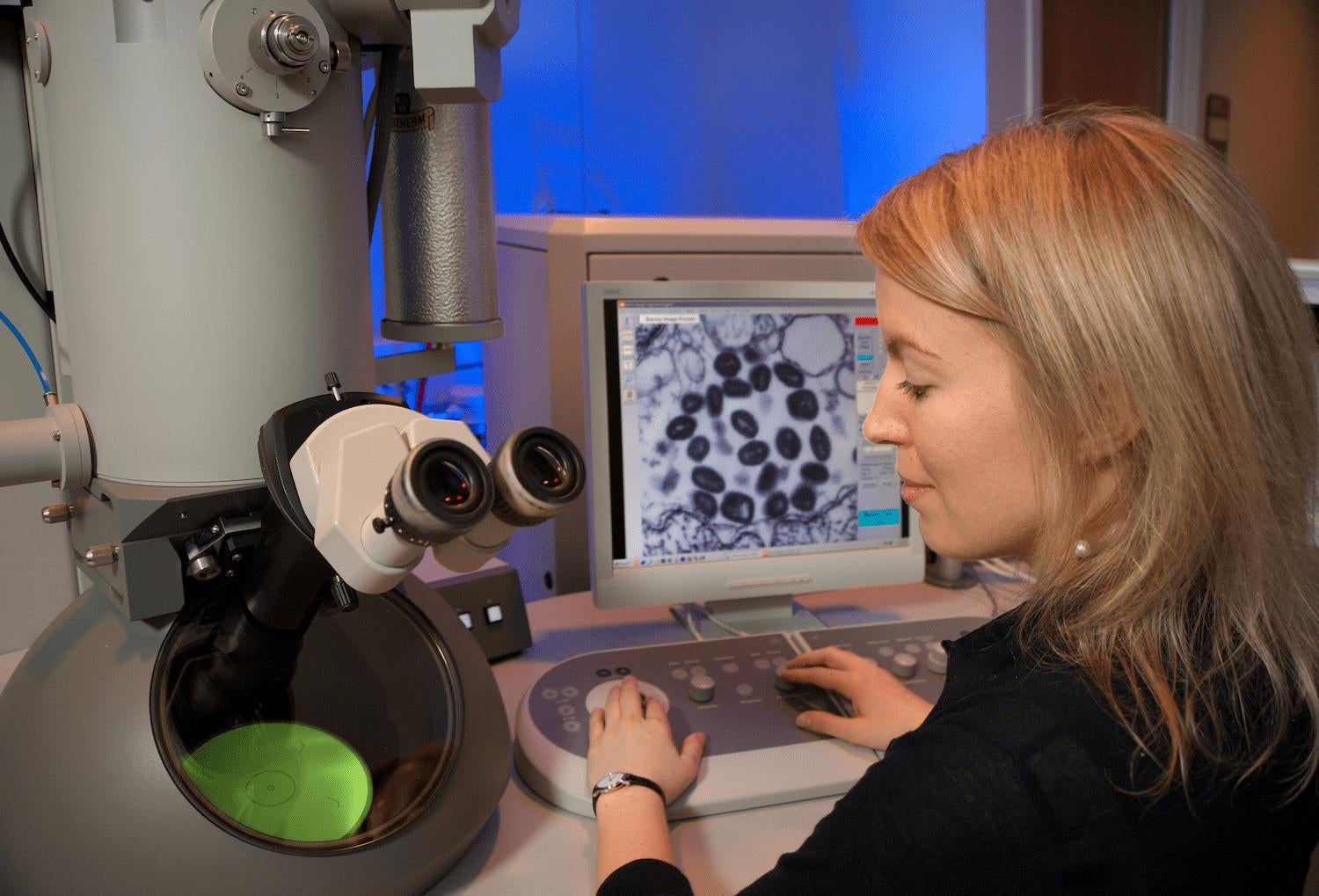
Working Principle of Electron Microscopy
Electron microscopes use
signals from the interaction of an electron beam with the sample to obtain
information about structure, morphology, and composition.
There are some basic steps involved in all EMs:
- A stream of high voltage electrons (usually 5-100 KeV) is emitted by the Electron Source (usually a heated tungsten or field emission filament) and accelerated in a vacuum toward the specimen using electric potentials.
- This stream is confined into a thin, monochromatic, focused electron beam using electromagnetic lenses.
- This electron beam is directed onto the sample using magnetic lenses.
- Interactions occur with the irradiated sample, affecting the primary electron beam and generating products such as secondary electrons or characteristic X-rays.
- Products of these interactions are detected and transformed into an image.
As simple as that!
Types of electron microscopes
We can distinguish two main
types of electron microscopes that use different techniques to obtain an
image.
Scanning Electron Microscope (SEM)
A scanning electron microscope produces
images of a sample by scanning the surface with a focused beam of electrons.
How exactly does it work? The electron beam interacts with atoms in the sample
and produces various signals containing information about the surface
topography and composition of the sample. The electron beam is scanned in a
raster scan pattern, and the position of the beam is combined with the
intensity of the detected signal to produce an image.
Because of its great depth of
focus, a scanning electron microscope is the EM analogue of a stereo light
microscope.
Transmission Electron Microscope (TEM)
Transmission Electron Microscope uses
a beam of electrons transmitted through an ultrathin specimen to form an image.
An image is formed as a result of the interaction of the electron beam with the
sample as the beam goes through the specimen. Using similar optics geometry as
in light microscopes the image gets magnified and focused on a series of
detectors.
A transmission electron
microscope can capture incredible details - even crystal structures with atomic
resolution, which is thousands of times smaller than objects visible through a
light microscope.
Transmission electron microscopy
can be used across different areas, such as the physical, chemical and
biological sciences. TEMs find application in cancer research,
virology, materials science as well as pollution, nanotechnology, semiconductor
research, and even palaeontology and palynology.
And there's more! Transmission Electron Microscopes can be used in scanning mode (STEM - Scanning Transmission Electron Microscope), combining both techniques’ advantages.


Comments
Post a Comment