Atomic Force Microscope
Atomic Force Microscope:
Principle, Parts, Uses
The atomic force microscope (AFM)
is a type of scanning probe microscope whose primary roles include measuring
properties such as magnetism, height, friction. The resolution is measured in a
nanometer, which is much more accurate and effective than the optical
diffraction limit. It uses a probe for measuring and collection of data
involves touching the surface that has the probe. An image is formed when the
scanning probe microscope raster-scans the probe over a section of the sample,
measuring its local properties concurrently. They also have piezoelectric
elements, which are electric charges that accumulate in selected solid
materials like DNA, biological proteins, crystal, etc, to enable tiny
accurate and precise movement during scanning upon an electric command. The
Atomic Force Microscope was invented in 1982, by scientists working in IBM,
just after the invention of the Scanning tunneling Microscope in 1980 by Gerd
Binnig and Heinrich Rohler by IBM Research in Zurich. That is when Binnig later
invented the Atomic Force Microscope, and it was first used experimentally in
1986. It was put on the market for commercial sale in 1989.
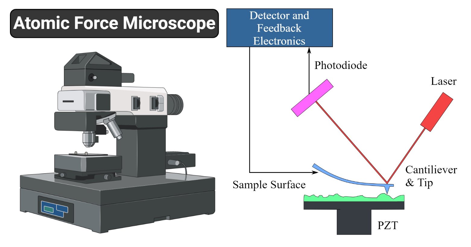
Figure:
Atomic Force Microscope (AFM). Source: Sagar Aryal and Wikipedia.
Principle
of Atomic Force Microscope
The Atomic Force Microscope works on the
principle of measuring intermolecular forces and sees atoms by using probed
surfaces of the specimen in a nanoscale. Its functioning is enabled by three
of its major working principles which include Surface sensing, Detection, and
Imaging.
- The Atomic Force Microscope (AFM) performs surface sensing by using a cantilever (an element that is made of a rigid block like a beam or plate, that attaches to the end of support, from which it protrudes making a perpendicularly flat connection that is vertical like a wall). The cantilever has a sharp tip that scans over the sample surface, by forming an attractive force between the surface and the tip when it draws closer to the sample surface. When it draws very close making contact with the surface of the sample, a repulsive force gradually takes control making the cantilever avert from the surface.
- During the deflection of the cantilever away from the sample surface, there is a change in the direction of reflection of the beam, and a laser beam detects the aversion, by reflecting off a beam from the flat surface of the cantilever. Using a positive-sensitive photo-diode (PSPD- a component that is based on silicon PIN diode technology and is used to measure the position of the integral focus of an incoming light signal), it tracks these changes of deflection and change in direction of the reflected beam and records them.
- The Atomic Force Microscope (AFM) takes the image of the surface topography of the sample by force by scanning the cantilever over a section of interest. Depending on how raised or how low the surface of the sample is, it determines the deflection of the beam, which is monitored by the Positive-sensitive photo-diode (PSDP). The microscope has a feedback loop that controls the length of the cantilever tip just above the sample surface, therefore, it will maintain the laser position thus generating an accurate imaging map of the surface of the image.
Amber Alert Accidentally Goes
Out Featuring Chucky the Killer Doll
Parts
of Atomic Force Microscope
Atomic Force Microscopes have several
techniques for measuring force interactions such as van der Waals, thermal,
electrical and magnetic force interactions for these interactions done by the
AFM, it has the following parts that assist in controlling its functions.
- Modified tips which are used to detect the sample surface and undergo deflections
- Software adjustments used to image the samples.
- Feedback loop control – they control the force interactions and the tip positions using a laser deflector. the laser reflects from the back of the cantilever and the tip and while the tip interacts with the surface of the sample, the laser’s position on the photodetector is used in the feedback loop for tracking the surface of the sample and measurement.
- Deflection – The Atomic Force Microscope is constructed with a laser beam deflection system. The laser is reflected from the back of the AFM lever to the sensitive detector. They are made from silicon compounds with a tip radius of about 10nm.
- Force measurement – the AFM works and depends highly on the force interactions, they contribute to the image produced. The forces are measured by calculation of the deflection lever when the stiffness of the cantilever is known. This calculation is defined by Hooke’s law, defined as follows:
F= -kz,
where F is the force, k is the stiffness of the lever, and z is the distance
the lever is bent.
Applications
of Atomic Force Microscope
This type of microscopy has been used in
various disciplines in natural science such as solid-state physics,
semiconductor studies, molecular engineering, polymer chemistry, surface
chemistry, molecular biology, cell biology, medicine, and physics.
Some of these applications include:
- Identifying atoms from samples
- Evaluating force interactions between atoms
- Studying the physical changing properties of atoms
- Studying the structural and mechanical properties of protein complexes and assembly, such as microtubules.
- used to differentiate cancer cells and normal cells.
- Evaluating and differentiating neighboring cells and their shape and cell wall rigidity.
Advantages
of Atomic Force Microscope
- Easy to prepare samples for observation
- It can be used in vacuums, air, and liquids.
- Measurement of sample sizes is accurate
- It has a 3D imaging
- It can be used to study living and nonliving elements
- It can be used to quantify the roughness of surfaces
- It is used in dynamic environments.
Disadvantages
of Atomic Force Microscope
- It can only scan a single nanosized image at a time of about 150x150nm.
- They have a low scanning time which might cause thermal drift on the sample.
- The tip and the sample can be damaged during detection.
- It has a limited magnification and vertical range.
References
and Sources
- https://www.first-sensor.com/en/products/optical-sensors/detectors/position-sensitive-diodes-psd/
- http://nanoscience.gatech.edu/zlwang/research/afm.html
- https://www.sciencedirect.com/topics/nursing-and-health-professions/scanning-probe-microscope
- https://www.researchgate.net/publication/256195163_The_Atomic_Force_Microscope
- https://quizlet.com/77735687/biology-campbells-chapter-12-cell-cycle-flash-cards/
- https://amedleyofpotpourri.blogspot.com/2018/09/atomic-force-microscopy.html
- http://www.eng.uc.edu/~beaucag/Classes/Characterization/ReflectivityLab/X-ray%20thin-film%20measurement%20techniques_V_X-ray%20reflectivity%20measurement.pdf
- http://www.edubilla.com/invention/atomic-force-microscopy/
- https://www.sciencedirect.com/science/article/pii/S0001868602000039
- https://www.researchgate.net/publication/304812262_Nanoscale_Imaging_of_RNA_with_Expansion_Microscopy
- https://www.coursehero.com/file/p45aldv/From-directly-above-youre-watching-a-fish-swim-183-m-beneath-the-surface-of-a/
- https://quizlet.com/35615640/a-certification-chapter-11-flash-cards/
- https://fas.org/man/dod-101/navy/docs/laser/fundamentals.htm
- https://aip.scitation.org/doi/10.1063/1.4931936


Comments
Post a Comment